what structure connects the right and left cerebral hemispheres?
 What Structure Connects The Right And Left Cerebral Hemispheres? - 99Science
BiopsychologyHemispheric brain Learning objectives The central nervous system (CNS), consists of the brain and spinal cord. BrainThe brain is a significantly complex organ composed of billions of interconnected neurons and glia. It is a bilateral or two-sided structure that can be separated into different lobes. Each lobe is associated with certain types of functions, but ultimately all areas of the brain interact with each other to provide the basis for our thoughts and behaviors. Column cords You can say that the spinal cord is what connects the brain to the outside world. That's why the brain can act. The spinal cord is like a relay station, but very smart. Not only does it run through messages to and from the brain, but it also has its own system of automatic processes, called reflexes. The upper part of the spinal cord is a set of nerves that merge with the brain trunk, where the basic processes of life, such as breathing and digestion, are controlled. In the opposite direction, the spinal cord ends just below the ribs — contrary to what we could expect, it does not extend to the base of the column. The spinal cord is functionally organized in 30 segments, corresponding to vertebrae. Each segment is connected to a specific part of the body through the peripheral nervous system. The nerves come out of the spine in each vertebrae. Sensory nerves bring messages; motor nerves send messages to muscles and organs. Messages travel from and to the brain through each segment. Some sensory messages are immediately performed by the spinal cord, without any input from the brain. Removed from a hot object and the knee shred are two examples. When a sensory message meets certain parameters, the spinal cord initiates an automatic reflex. The signal passes from the sensory nerve to a simple processing center, which initiates an engine command. The seconds are saved, because the messages don't have to go to the brain, be processed and be sent back. In survival, spinal reflexes allow the body to react extraordinarily quickly. The spinal cord is protected by bone vertebrae and glued in cerebrospinal fluid, but lesions still occur. When the spinal cord is damaged in a particular segment, all lower segments are cut from the brain, causing paralysis. Therefore, the minor in the damage of the column is, the least number of functions that an injured individual will lose. Neuroplicity Bob Woodruff, ABC reporter, suffered a traumatic brain injury after a bomb exploded next to the vehicle he was covering news in Iraq. As a result of these injuries, Woodruff experienced many cognitive deficits including memory and language difficulties. However, with time and with the help of intensive amounts of cognitive and speech therapy, Woodruff has shown an incredible recovery of the function (Fernandez, 2008, October 16). One of the factors that made this recovery possible was neuroplasticity. Neuroplicity refers to how the nervous system can change and adapt. Neuroplicity can occur in various ways, including personal experiences, development processes or, as in the case of Woodruff, in response to some kind of damage or injury that has occurred. Neuroplicity can involve the creation of new sinapsis, the sinapsis pod that is no longer used, changes in glyal cells, and even the birth of new neurons. Because of neuroplicity, our brains are constantly changing and adapting, and while our nervous system is more plastic when we are very young, as Woodruff suggests, it is still capable of significant changes later in life. Two hemispheres The surface of the brain, known as the cerebral cortex, is very unequal, characterized by a distinctive pattern of folds or strokes, known as gyri (singular: spin), and the grooves, known as sulci (singular: sulcus), shown in Figure 1. These spins and sulci form important milestones that allow us to separate the brain into functional centers. The most prominent sulcus, known as the longitudinal fissure, is the deep groove that separates the brain in two halves or hemispheres: the left hemisphere and the right hemisphere. Figure 1. The surface of the brain is covered with gyri and sulci. A deep sulcus is called fisura, like the longitudinal fissure that divides the brain into left and right hemispheres. (credit: work modification by Bruce Blaus) There is evidence of specialization of the function, which is referred to as lateralization, in each hemisphere, mainly regarding differences in linguistic functions. The left hemisphere controls the right half of the body, and the right hemisphere controls the left half of the body. Decades of research on the lateralization of function by Michael Gazzaniga and his colleagues suggest that a variety of functions ranging from causal and effective reasoning to self-recognition can follow patterns that suggest a certain degree of hemispheric domain (Gazzaniga, 2005). For example, it has been shown that the left hemisphere is superior to form associations in memory, selective attention and positive emotions. The right hemisphere, on the other hand, has proved to be superior in the perception of the field, excitement and negative emotions (Eyret, 2006). However, it should be noted that research on which hemisphere is dominant in a variety of different behaviors has produced inconsistent results, and therefore it is probably better to think about how the two hemispheres interact to produce a given behavior rather than attribute certain behaviors to one hemisphere versus the other (Banich & Heller, 1998). The two hemispheres are connected by a thick band of neuronal fibers known as the callosum body, which consists of about 200 million axons. The callosum body allows the two hemispheres to communicate with each other and allows the information to be processed on one side of the brain to be shared with the other side. Normally, we are not aware of the different roles that our two hemispheres play in everyday functions, but there are people who know well the capabilities and functions of their two hemispheres. In some cases of severe epilepsy, doctors choose to separate the callosum body as a means of controlling the spread of seizures (Figure 2). While it is an effective treatment option, it results in individuals who have "multiplied brains". After surgery, these brain patients divided show a variety of interesting behaviors. For example, a separate brain patient cannot name an image shown in the patient's left visual field because the information is only available in the non-verbal right hemisphere. However, they are able to recreate the image with their left hand, which is also controlled by the right hemisphere. When the most verbal left hemisphere sees the image that the hand drew, the patient is able to name it (assuming that the left hemisphere can interpret what was drawn by the left hand). Figure 2. (a, b) The callosum body connects the left and right hemispheres of the brain. c) A scientist separates this brain from spread sheep to show the callosum body between the hemispheres. (credit c: modification of work by Aaron Bornstein) Much of what we know about the functions of the different areas of the brain comes from studying changes in the behavior and capacity of individuals who have suffered damage to the brain. For example, researchers study behavioral changes caused by strokes to learn about the functions of specific brain areas. A stroke, caused by an interruption of blood flow to a region of the brain, causes a loss of brain function in the affected region. The damage may be in a small area, and if it is, this gives researchers the opportunity to link any resulting behavioral change to a specific area. The types of deficits shown after a stroke will depend to a large extent on where the damage occurred in the brain. Consider Theona, a smart, self-sufficient woman, who is 62 years old. Recently, he suffered a stroke on the front of his right hemisphere. As a result, you have a great difficulty moving your left leg. (As you learned earlier, the right hemisphere controls the left side of the body; also, the main motor centers of the brain are in the front of the head, in the frontal lobe.) Theona has also experienced behavioral changes. For example, while in the grocery store product section, you sometimes eat grapes, strawberries and apples directly from your containers before paying for them. This behavior—which would have been very embarrassing for her before the stroke—is consistent with damage to another region in the frontal lobe—the prefrontal cortex, which is associated with judgment, reasoning and impulse control. See ItWatch this video to see an incredible example of the challenges faced by a brain patient split shortly after surgery to see your body callosum. You can. See this second video about another patient who suffered dramatic surgery to prevent her seizures. You will learn more about the ability of the brain to change, adapt and reorganize, also known as brain plasticity. You can. Try it. GlossaryContribute!

What Structure Connects The Right And Left Cerebral Hemispheres? - 99Science

What do you call that part of the Brain that connects the right and the left hemispheres? - Quora

Brain Hemispheres | Introduction to Psychology
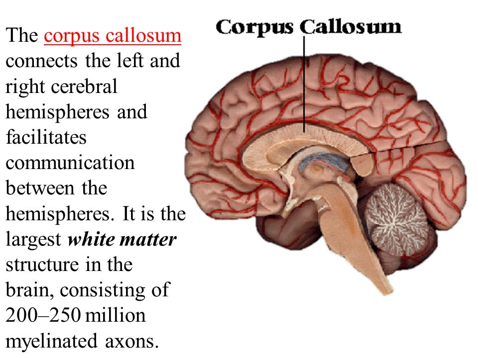
The Brain The brain is composed of the cerebrum, cerebellum, and brainstem. - ppt video online download
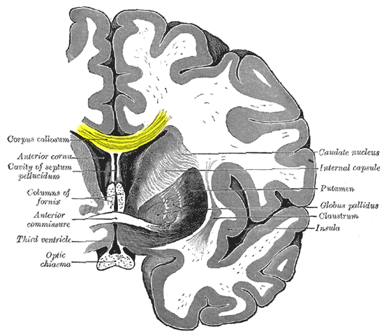
What connects the two hemispheres of the cerebrum? | Socratic

What do you call that part of the Brain that connects the right and the left hemispheres? - Quora

Cerebral hemisphere | anatomy | Britannica

Mastering A&P Chapter 12 - The Central Nervous System Flashcards | Quizlet
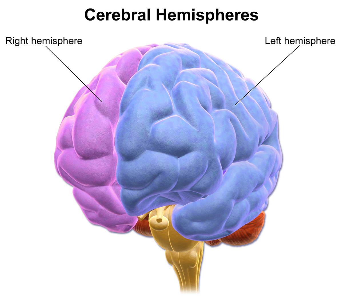
Cerebral hemisphere - Wikipedia

Mastering A&P Chapter 12 - The Central Nervous System Flashcards | Quizlet

What do you call that part of the Brain that connects the right and the left hemispheres? - Quora

The Brain Hemispheres: Left Brain/Right Brain Communication and Control - Anatomy and Physiology Class (Video) | Study.com
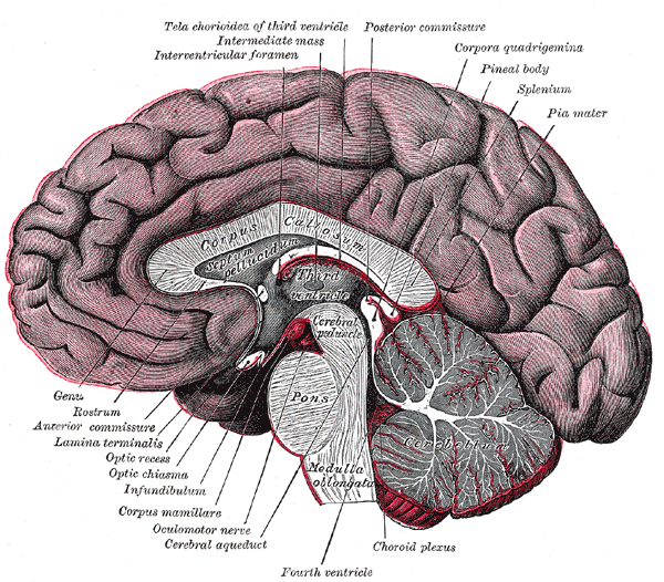
Which portion of the cerebellum connects the right and left cerebellar hemispheres? | Socratic

Mastering A&P Chapter 12 - The Central Nervous System Flashcards | Quizlet

Left and Right Hemisphere of the Brain | Functions & Characteristics

The Cerebrum and Cerebral Hemispheres

Mastering A&P Chapter 12 - The Central Nervous System Flashcards | Quizlet

Solved: QUESTION 25 The Right And Left Cerebral Hemisphere... | Chegg.com

The Brain Hemispheres: Left Brain/Right Brain Communication and Control - Anatomy and Physiology Class (Video) | Study.com
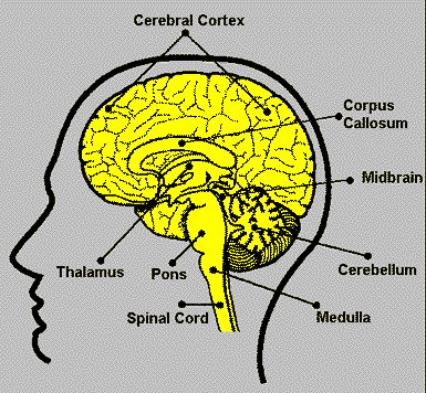
Neuroscience Resources for Kids
/Corpus-Callosum-58d16cd45f9b581d727aa487.jpg)
Corpus Callosum and Brain Function

The Cerebrum | Boundless Anatomy and Physiology

Right Brain vs. Left Brain: Language, Dominance, and Their Shared Roles

Know your brain: Corpus Callosum — Neuroscientifically Challenged

Split-brain syndrome explained | Britannica

Cerebral hemisphere - Wikipedia
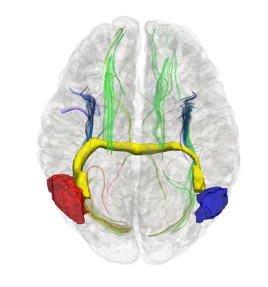
People Missing Brain Wiring Form Unique Neural Connections | Live Science
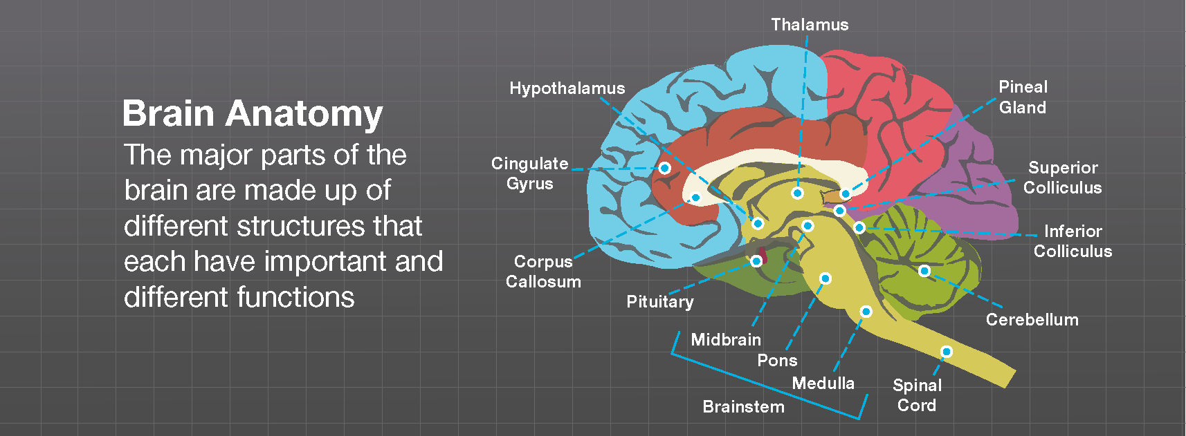
Brain Anatomy - Brainlab.org

The Cerebrum | Boundless Anatomy and Physiology

The brain
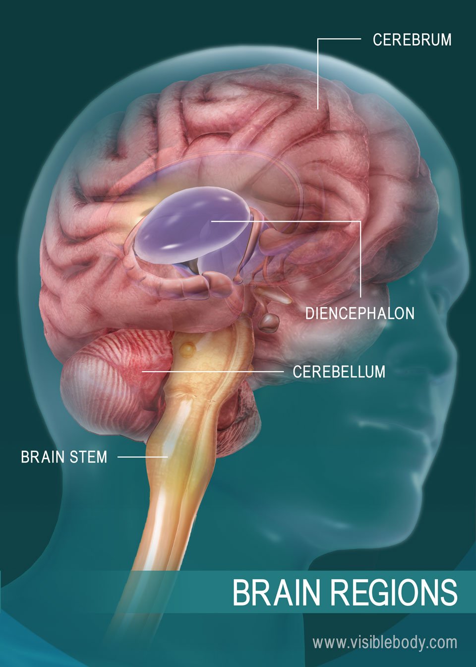
The Human Brain
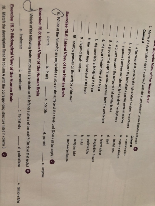
Solved: Superior View Of The Human Brairn 7. Match The Des... | Chegg.com
/LeftvsRight-01-5a206273ec2f64003722380d.png)
Left Brain vs. Right Brain Dominance

Cerebral hemisphere - Wikipedia

12.3: Cerebrum - Medicine LibreTexts
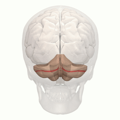
Cerebellum - Physiopedia

What structure connects the right and left cerebral hemispheres? | Study.com

The right side of the brain Visual Imagery Music What structure connects the | Course Hero
/brain_spinal_cord-57fe96b15f9b5805c26d5072.jpg)
The Central Nervous System in Your Body
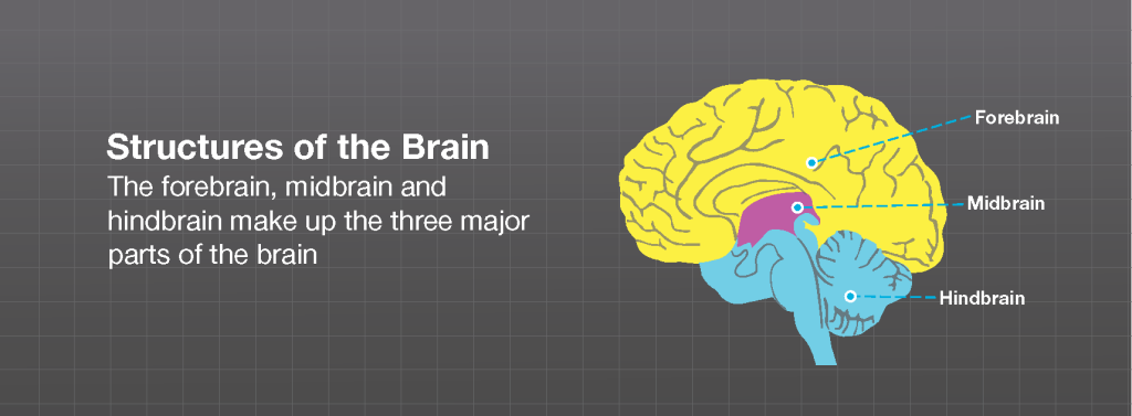
Brain Anatomy - Brainlab.org
Posting Komentar untuk "what structure connects the right and left cerebral hemispheres?"